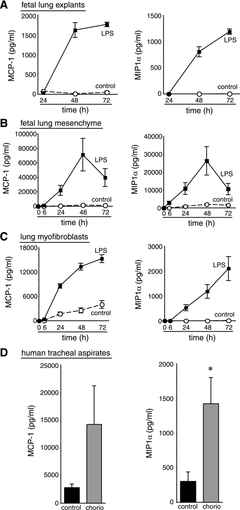
Fig. 6.
LPS stimulates release of MIP-1α and MCP-1. Fetal mouse lung explants (A), primary fetal mouse lung mesenchymal cells (B), and mouse lung myofibroblasts (C) were cultured in control media or in the presence of E. coli LPS (250 ng/ml). Media were collected at indicated time points, and chemokine concentrations were measured by Luminex assay. Each sample was measured in triplicate. D: MCP-1 and MIP-1α were detected in the tracheal aspirate fluid of newborn preterm infants exposed to maternal chorioamnionitis (chorio) by SearchLight assay (*P < 0.05; 7 control and 9 chorioamnionitis samples tested). Error bars ± standard error of the mean.

