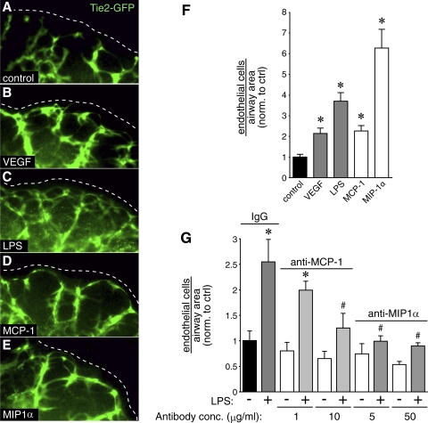
Fig. 7.
MCP-1 and MIP-1α stimulate vascular development in fetal mouse lung explants. A–E: saccular stage fetal lung explants from Tie2-GFP mice were cultured in the presence of VEGF, LPS, MCP-1, or MIP-1α. F: after obtaining confocal images of the Tie2-GFP explants, the number of endothelial cells surrounding each saccular airway was measured, corrected for airway area, and compared with control. (*P < 0.001; n = 12). G: antibodies against MCP-1 and MIP-1α inhibit LPS-mediated angiogenesis. Nonimmune rat IgG included as control. (*P < 0.05 compared with nonimmune IgG, #P < 0.05 compared with LPS with nonimmune IgG; n = 7). Error bars ± standard error of the mean. conc., Concentration.

