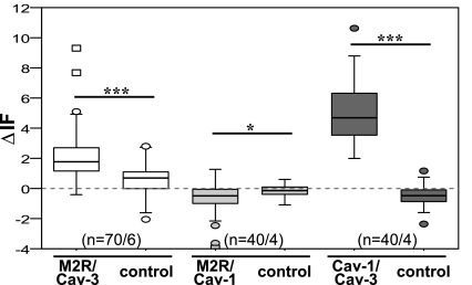
Fig. 4.
Detection of close spatial association of M2R/Cav-3 and Cav-1/Cav-3 by double-labeling indirect immunofluorescence and CLSM-fluorescence resonance energy transfer (FRET) analysis in airway smooth muscle of murine bronchi in situ. Changes in donor fluorescence (ΔIF) in the membrane area of bronchial smc in experimental compared with control group are shown. For M2R/Cav-3 and Cav-1/Cav-3, ΔIF is higher in experimental groups than in controls. ***P ≤ 0.001, *P ≤ 0.01; n = number of regions of interest/animals. Box plots: percentiles 0, 25, median, 75, 100; extreme values (○), outlier (☐).

