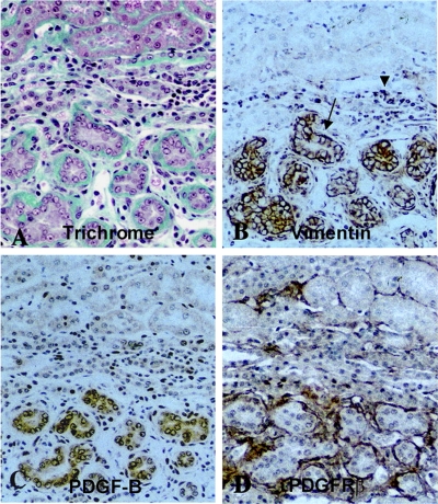
Fig. 5.
Sections from kidneys of rats microembolized through the left renal artery with 20- to 30-μm-diameter acrylic microspheres and examined 4 wk later. Serial paraffin sections of methyl Carnoy's fixed tissue were stained with Masson's trichrome (A) or immunohistochemically stained for vimentin (B), PDGF-B (C), or PDGF receptor (PDGFR)-β (D). Original magnification ×200. Reproduced from Ref. 144 with kind permission from Dr. Akira Hishida, first department of Medicine, Hamamatsu University School of Medicine, Hamamatsu, Japan. Reproduced from Am J Pathol 158: 75–85, 2001; with permission from the American Society for Investigative Pathology.

