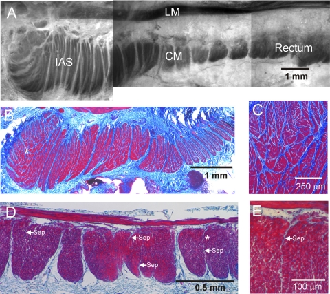
Fig. 1.
Morphological features of the monkey rectoanal region. A: composite image of a thick cross section from the rectoanal region; smooth muscle is contrasted with toluidine blue staining. The longitudinal (LM) and circular (CM) muscle layers can be seen separated by connective tissue. Prominent septal structures (white) are also apparent extending from the myenteric to the submucosal edge of the circular muscle separating this layer into bundles. The pinning procedure used for this image has exaggerated the gap between longitudinal and circular muscle layers. A more representative separation can be seen in D. B–E: thin (3 μm) cross sections of the internal anal sphincter (IAS) and rectum stained with Masson's trichrome to visualize both smooth muscle (red/purple) and connective tissue (blue). B: low magnification image of the IAS again showing prominent septal structures extending from myenteric (top) to submucosal (bottom) edge. Additional septal structures are apparent further separating the muscle into “minibundles.” C: higher magnification image of the region marked with an asterisk in B showing numerous minibundles (red) separated by connective tissue septa (blue). D: lower magnification image of rectum showing division of the circular muscle layer into compact bundles extending from the myenteric to the submucosal edge and separated by connective tissue septa. A few additional smaller septa can be seen penetrating for variable distances into muscle bundles (arrows labeled “sep”). The region marked with an asterisk in D is shown at higher magnification in E. Cells can be seen between longitudinal and circular muscle layers and these likely include both neurons and interstitial cells of Cajal (ICC) of the myenteric plexus.

