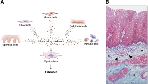
Fig. 3.
A: working model for esophageal fibrosis. Chronic inflammation can drive fibrogenesis. This process involves essentially all cell types that can contribute to the activation of local mesenchymal cells. B: Masson trichrome staining of a peptic esophageal stricture. Collagen fibers are depicted in blue. Massive subepithelial and submucosal collagen accumulation (*) with neoangiogenesis and an inflammatory infiltrate (arrows) can be noted. Magnification ×10.

