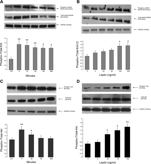
Fig. 5.
A–B: leptin activates ERK1/2 isoforms of MAPK. T84 cell monolayers were cultured in serum-free media (DMEM/F12) for 24 h followed by exposure to recombinant human leptin. Leptin was added at 100 ng/ml for the times indicated (A) or at different doses for 5 min (B). Cellular extracts were fractioned by 12% SDS-PAGE, and Western immunoblotting was performed with a rabbit polyclonal anti-phospho-p44/42 MAPK and anti-total ERK as described in materials and methods. C–D: leptin activates Akt in T84 cells. Monolayers were cultured in serum-free media (DMEM/F12) for 24 h followed by exposure to human recombinant leptin at a concentration of 100 ng/ml for up to 15 h (C) or at different doses for 5 min (D). Cellular extracts were analyzed by Western immunoblotting with anti-phospho-Akt 1/PKB-α and anti-total Akt as described in materials and methods. Equal loading was also assessed using a rabbit monoclonal antibody to GAPDH. In all panels, representative blots are shown above with summary data from at least 3 similar experiments, analyzed by densitometry, below. These latter values are means ± SE, and those that differ significantly from controls are designated with asterisks (*P < 0.05; **P < 0.01 by ANOVA).

