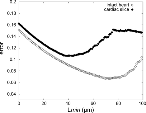
Fig. 5.
Error bounds estimate for the intact heart (◊) and cardiac slice (♦) preparations. The magnitude of the error is calculated based on the no. of actual measurements, available in a given data set, with longest line perpendicular to the striation patterns within cell boundaries (L) ≥ minimum cell length in the image plane (Lmin).

