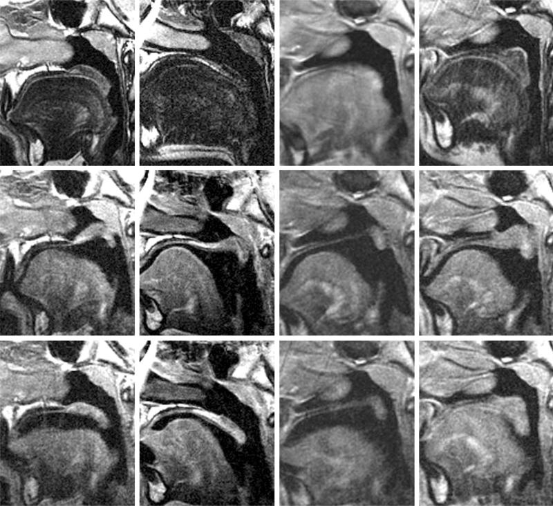
Fig. 1.
Velopharyngeal mechanism visualized on magnetic resonance imaging. (Above) Velum at rest during nasal breathing. (First column) Control child 1; (second column) control child 2; (third column) submucous cleft palate child preoperatively; (fourth column) submucous cleft palate child postoperatively. The velum rests passively on the tongue during nasal breathing, maintaining airflow through the nasopharynx. (Center) Velum during phonation of “e” sound, demonstrating closure of the nasopharynx. (First column) Control child 1; (second column) control child 2; (third column) submucous cleft palate child preoperatively; (fourth column) submucous cleft palate child postoperatively. Note the absence of muscle in the velum of the submucous cleft palate patient preoperatively, with a persistent velopharyngeal gap on phonation (third column) compared with considerable muscle bulk in this location postoperatively with closure of the velopharyngeal gap (fourth column). (Below) Velum during phonation of “n” sound, with airflow through both the nasal and oral cavities. (First column) Control child 1; (second column) control child 2; (third column) submucous cleft palate child preoperatively; (fourth column) submucous cleft palate child postoperatively. The velum rests in an intermediate position, with airflow through both the nasopharynx and the oropharynx.

