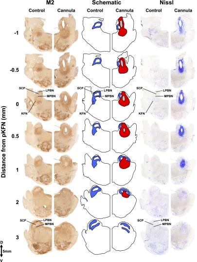
Fig. 3.
Histochemical and immunohistochemical staining for Nissl substance and M2 receptors of hemisections from 1 control goat and 1 goat with cannula implanted into the KFN. These hemisections illustrate the rostral-caudal changes in the LPBN and MPBN and KFN beginning 1 mm caudal to the peak in number of KFN neurons and extent to 3 mm rostral to the peak. Blue shaded area illustrates the orientation of the LPBN, MPBN, and KFN relative to the SCP. The tract of the cannula (in gray) extends over 2 mm, and the area of devoid of neurons (in red) extends over 3 mm in the rostral-caudal direction. Note the clear presence of the KFN at its peak number of neurons (0 mm) and its absence at a more rostral distance (3 mm).

