Fig. 3.
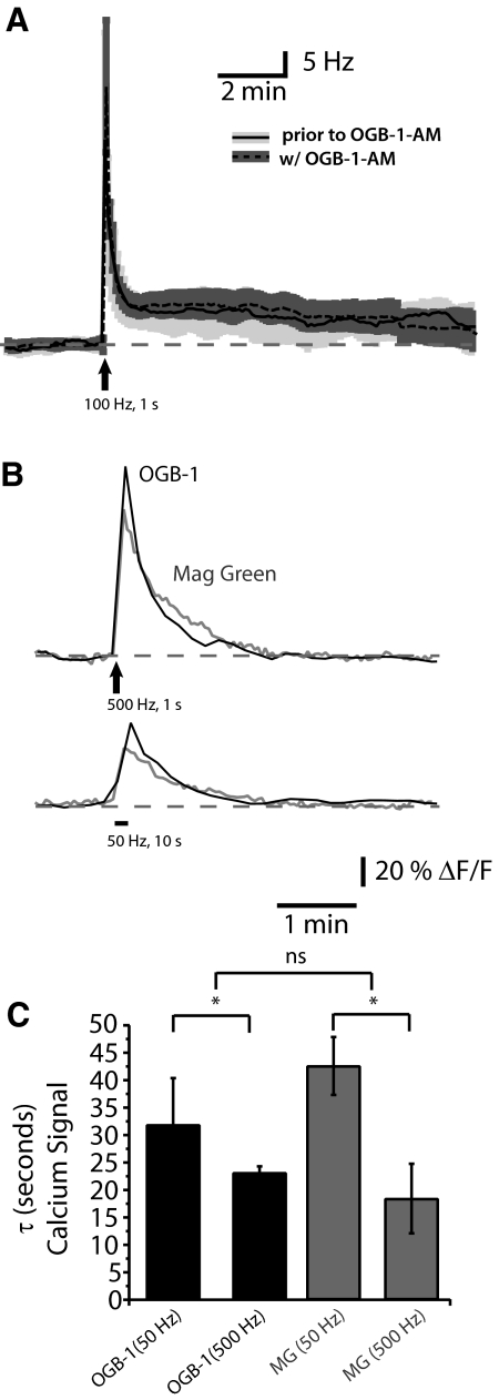
Dye loading does not alter PMn activity. A: LTFE (Hz) in response to afferent fiber stimulation (100 pulses at 100 Hz) prior to dye loading (average: solid black, SE: light gray) and with dye loaded into the PMn neurons (average: dashed black, SE, dark gray). B: changes in fluorescence in response to synaptic stimulation (500 pulses at 500 Hz or 500 pulses at 50 Hz) of high affinity calcium-binding dye (Oregon Green BAPTA-1, in black), the low affinity calcium-binding dye magnesium green (Kd = 6 μM; ΔF/F, in gray). C: decay kinetics of the Ca2+ signal when using OGB-1 (Kd ∼205 nM) vs. magnesium green. Decay kinetics (τ) were compared after synaptically stimulating the PMn with 500 pulses at 50 Hz and 500 Hz [ANOVA F(3,29) = 8.604, P < 0.01]. No significant difference between the decays of the Ca2+ signal using different dyes (Bonferonni's multiple comparisons), but significant difference was detected between 50 and 500 Hz stimulations (Bonferonni's multiple comparisons, P < 0.05).

