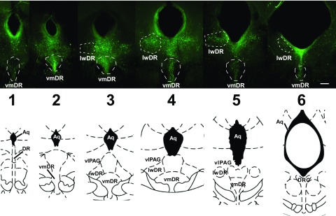
Fig. 1.
5-Hydroxytryptamine (5-HT, serotonin) neurons extend across the rostrocaudal extent of the dorsal raphe (DR). Top panel: images of 200 μm thick slices across the rostrocaudal extent of the DR following immunohistochemical detection of tryptophan hydroxylase. White dotted lines indicate the regions targeted for electrophysiology (scale bar: 208 μm). Bottom panel: corresponding brain atlas images (Paxinos and Watson 1996). Numbers indicate rostrocaudal level used for correlative analysis. Aq, aqueduct; DRC, dorsal raphe, caudal part; lwDR, lateral wing of the DR; vlPAG, ventrolateral periaqueductal gray; vmDR, ventromedial DR.

