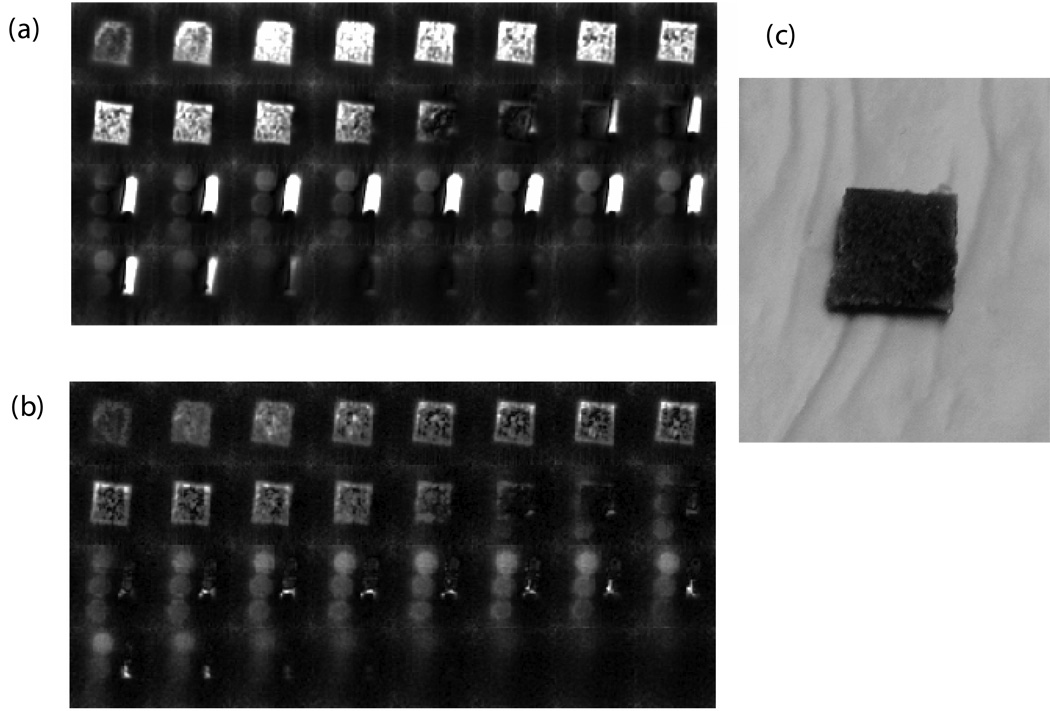
Figure 5. SMRI and WASPI images of a trabecular bone specimen, marrow specimen, and polymer phantoms and photograph of the trabecular bone specimen.
MRI imaging of bone specimens were taken along with the marrow specimen and three polymer pellets. a) SMRI (no water and fat suppression). b) WASPI. c) Photograph of the trabecular specimen.

