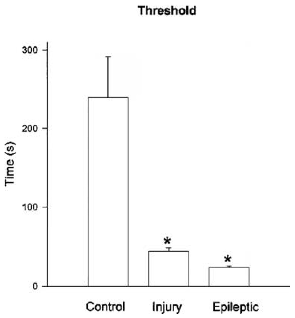
FIG. 4.
Histogram quantitating differences in status epilepticus (SE) thresholds. Note the statistically significant differences in SE afterdischarge duration threshold between control, brain-injured, and epileptic hippocampal–entorhinal cortical (HEC) slices. Control slices had significantly higher thresholds than either brain-injured or epileptic HEC slices *(p < 0.0001, t test), and brain-injured slices had SE thresholds significantly longer than epileptic slices *(p < 0.005, t test).

