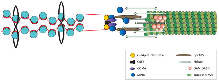
Figure 4.
A schematic representation of the interface between kinetochore microtubule, kinetochore and pericentric chromatin. The microtubule ( green, right) is a 25-nm tubule comprised of 13 protofilaments. Pericentric chromatin (blue nucleosomes and red DNA) is organized into an intramolecular loop in mitosis. The dimensions of a single nucleosome are 5 × 11.5 nm. The dimension of an intramolecular loop would be approximately 23 nm. The two major polymers (nucleosomal DNA and microtubules) are similar in cross-sectional dimension. The kinetochore is a proteinaceous structure linking these two polymers in mitosis.

