Fig. 1.
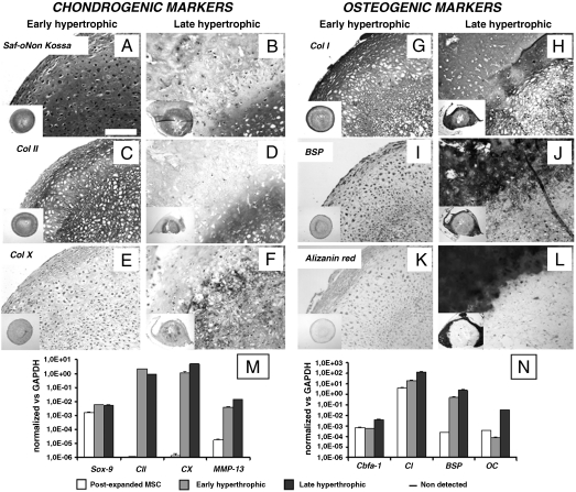
In vitro maturation of hypertrophic cartilage tissues engineered from human adult MSC. In vitro culture conditions determined the composition and structure of the tissues generated. (A, C, E, and G) Early hypertrophic samples displayed a cartilaginous ECM rich in GAG and Col II with deposition of Col X and Col I in defined regions. (I and K) In the periphery of early hypertrophic samples, low BSP levels were detected, but no calcium was deposited. (B, D, F, H, J, and L) Late hypertrophic samples underwent further maturation in vitro and developed two distinct regions: an inner hypertrophic core (B, D, and F) rich in GAG, Col II, and Col X, and an outer mineralized rim (B, H, J, and L) with a high mineral content, Col I, and BSP. All pictures were taken at the same magnification. (Scale bar: 200 μm.) The insets display low magnification overviews of the entire tissues. (M and N) Quantitative real-time RT-PCR demonstrated an up-regulation of hypertrophic (Col X and MMP-13) and osteogenic (cbfa-1, OC, BSP) markers when comparing late with early hypertrophic tissues. Postexpanded MSC remained in an undifferentiated state but expressed both SOX-9 and Cbfa-1 in combination with high type I collagen and low type II collagen levels.

