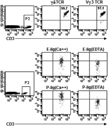
Fig. 1.
DETC in normal WT C57BL/6 mice express E-lig and P-lig. Epidermal cell suspensions prepared from 8-week-old WT mice were stained with fluorescence-conjugated anti-CD3, anti-γδ TCR, and anti-Vγ3 TCR, followed with analysis by flow cytometry. The numbers in quadrants indicate the percentages in CD3+ T cells. E- and P-lig expression was examined by incubating skin cells with rmCD62E/Fc or rmCD62P/Fc chimera in HBSS buffer containing 2 mM calcium. HBSS buffer supplemented with 5 mM EDTA was used for the controls. Data are representative of at least three independent experiments.

