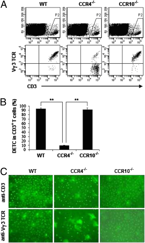
Fig. 7.
DETC are diminished in CCR4−/− but not in CCR10−/− mice. (A) Epidermal cells were prepared from 8-week-old WT, CCR4−/−, or CCR10−/− C57BL/6 mice. Cells then were washed thoroughly and labeled with fluorescence-conjugated anti-CD3 and anti-Vγ3 TCR mAbs for DETC analysis by flow cytometry. (B) Summary data on the percentages of DETC in CD3+ T cells. **, P < 0.01. (C) DETC on epidermal sheets were visualized by labeling with FITC-conjugated anti-CD3 or anti-Vγ3 TCR mAb (×100). One of three independent experiments with similar results is shown.

