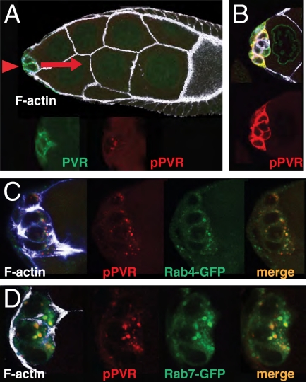
Fig. 2.
Detection of active PVR in border cells initiating migration. (A) Early stage-9 egg chamber, with border cells (arrowhead) initiating migration. In this and all subsequent images, direction of migration is to the right (red arrow), the genotype is slbo-Gal4 + UAS-PVR, and white/blue is phalloidin signal. Total PVR (green); pPVR (red) signals are shown separately in the enlargement of the border cell cluster below. (B) Border cells as in A but after incubation with the phosphatase inhibitor vanadate for 10 min. (C) Border cell cluster from females expressing Rab4-YFP ubiquitously (green), showing overlap with the pPVR signal (merge). (D) Border cell cluster from females carrying UAS-Rab7-GFP. Note that overexpression of Rab proteins may change the size of targeted (endosomal) compartments, disallowing quantitative comparisons between genotypes (see, e.g., ref. 27).

