
Figure 4.
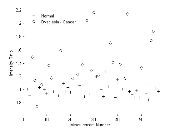
Threshold ratio of multi-spectral digital microscope developed by Roblyer [12]. Scatter plot of the normalized ratio of red to green MFI at 450 nm excitation with biopsy as gold standard using the multispectral digital microscope. The threshold value indicated by the horizontal line for differentiating premalignant lesions and cancer from normal sites is 1.09.