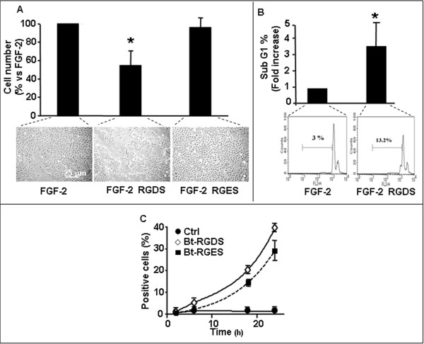
Figure 1.
SK-MEL-110 proliferation and apoptosis. A: SK-MEL-110 seeded on collagen-IV (50 μg/ml) were treated for 48 h with RGDS or RGES (500 μg/ml) in the presence of FGF-2 (10 ng/ml). Data are expressed as mean ± SD of 5 experiments carried out in duplicate. Representative images are reported. B: SK-MEL-110 apoptosis was quantified by FACS analysis of PI-stained cells after 48 h RGDS treatment (500 μg/ml) (p < 0.05 vs FGF-2). Three independent experiments were performed and quantified; one representative experiment is shown. C: Biotinylated-RGDS and RGES internalization in SK-MEL-110 was measured by FACS. Cells were treated for different time points (2, 6, 16 and 24 h) with bt-RGDS or bt-RGES; internalization was revealed by PE-avidin. Three independent experiments were performed.

