Fig. 4.
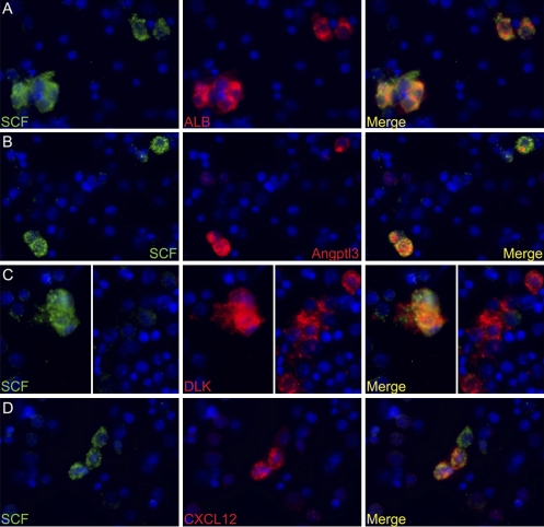
SCF+ cells in E15.5 fetal liver are also positive for ALB, Angptl3, and DLK expression but are heterogeneous for CXCL12 expression. (A) Double immunocytochemistry for SCF (green) and ALB (red) expression in total fetal liver cells. DAPI was used to stain nuclei (blue). SCF+ cells are also positive for ALB expression. (B) Double staining for SCF and Angptl3. Three SCF+ cells are shown, all of which are positive for Angptl3 expression. (C) Double staining for SCF and DLK. (Left) Cluster of SCF+ cells that are also DLK+. DLK is also expressed by other cells, as indicated (Right) by a group of DLK+ cells that are SCF–. (D) Only a fraction of SCF+ cells are also CXCL12+. Shown are three SCF+ cells, two of which are CXCL12+ and one is CXCL12– (white arrow).

