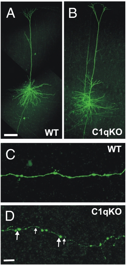
Fig. 3.
Increased density of axonal boutons in cortical layer V pyramidal neurons of the C1q KO mice. (A and B) Confocal images of biocytin filled layer V pyramidal neurons of C1q KO (A) and WT (B) mice. (C and D) A segment of the axon from the control cell (C) and C1q KO neuron (D). Large and small arrows in D point to examples of large (>1 μm) and small (≤1 μm) boutons, respectively. (Scale bars: A for A and B, 100 μm; D for C and D, 10 μm.)

