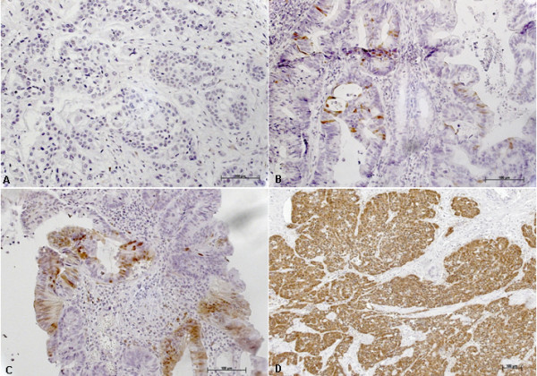
Figure 2.

Immunohistochemical analysis of p16INK4A expression in colorectal carcinomas. A: no p16INK4A expression (negative) in tumor cells. B: weak expression of p16 in tumor cells. C: moderate expression of p16INK4A in tumor cells and D: strong expression of p16INK4A with a strong intensity in tumor cells.
