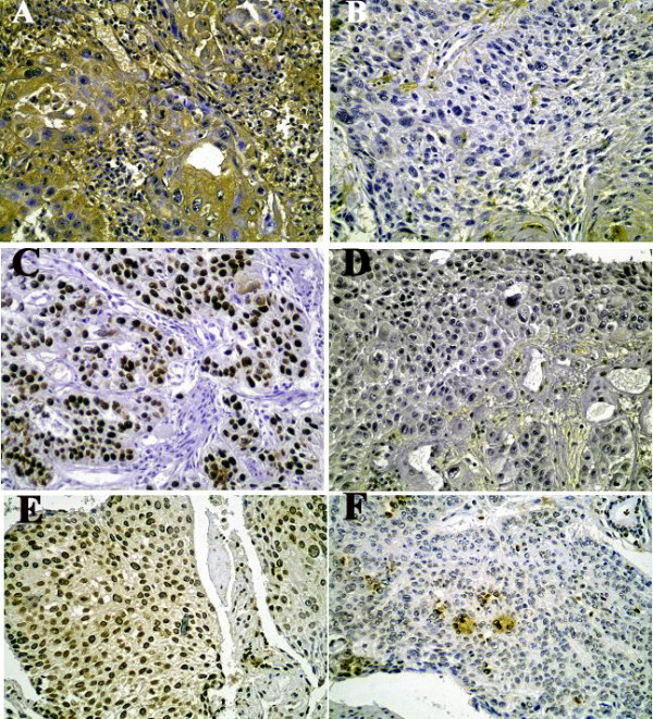
Figure 2.

Typical images of Immunohistochemical staining. a) Positive p16 immunoreactivity. b) Negative p16 immunoreactivity c) Overexpression of p53 protein. d) Negative p53 immunostaining e) Overexpression of MDM2 protein. f) Negative MDM2 immunoreactivity.
