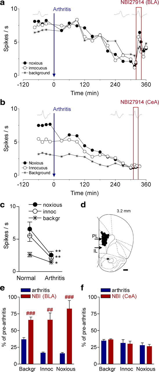
Figure 5.

Deactivation of BLA restores normal activity in PFC neurons. a, Time course of activity changes in an individual prelimbic mPFC neuron recorded continuously before and after induction of an arthritis pain state (extracellular single-unit recordings in an anesthetized rat). Each symbol shows background activity or responses to innocuous and noxious stimuli applied to the knee joint (see Fig. 1). Administration of a CRF1 receptor antagonist (NBI27914, 10 μm, concentration in microdialysis fiber; 20 min) into the BLA increased the activity of the mPFC neuron. b, Time course of activity changes in another prelimbic mPFC neuron recorded before and after arthritis induction. Administration of NBI27914 (10 μm) into the CeA had no effect. c, Significant decrease of background and evoked activity in the mPFC neurons 5–6 h after arthritis induction (n = 26 neurons). *p < 0.05, **p < 0.01 (compared with normal; paired t test). d, Recording sites in the prelimbic cortex. PL, Prelimbic part of the mPFC; IL, infralimbic part of the mPFC. e, Deactivation of the BLA with NBI27914 (10 μm) increased the background activity of mPFC neurons and their responses to innocuous and noxious stimuli significantly (n = 5 neurons; p < 0.01–0.001, paired t test). f, Administration of NBI27914 into the CeA had no effect on mPFC neurons (n = 5). e, f, Bar histograms show mean ± SE normalized to control values before arthritis (set to 100%). For each neuron, two to three data points were averaged before arthritis, 5–6 h after induction of arthritis, and during drug application (>15 min). ##,###p < 0.01, 0.001 (compared with predrug; paired t test).
