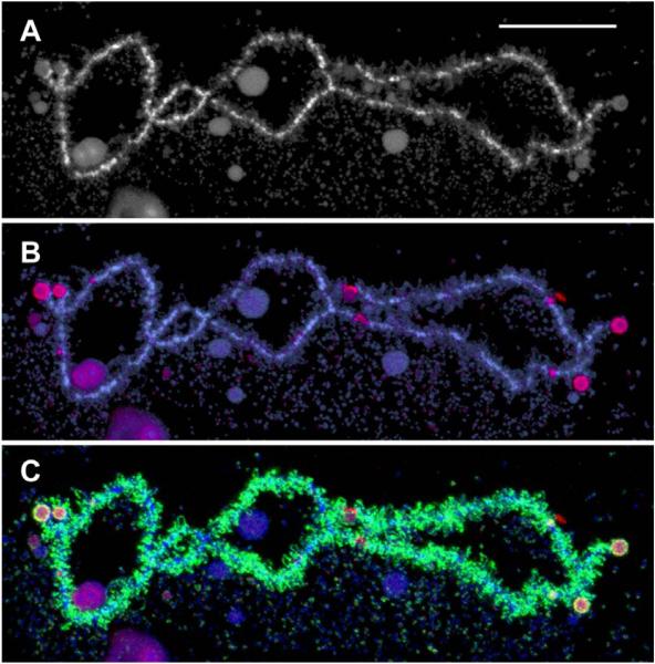
Figure 7.
A single LBC from a mature GV of X. tropicalis. An advantage of X. tropicalis over X. laevis for the study of LBCs is the smaller chromosome number (n = 10 vs n = 18) and the fact that the genome has been sequenced (http://www.xenbase.org). Working maps of the LBCs have also been published recently [35]. A. The longest chromosome of the set, stained with the DNA-specific dye DAPI. B. Immunofluorescent staining (red) with an antibody that recognizes RNA polymerase III. A few interstitial sites of pol III transcription are evident. The nature of the stained spherical objects on the chromosome ends is not known. Counterstained with DAPI (blue). C. The same chromosome showing staining for pol II (green) and pol III (red). The majority of loops are transcribed by pol II. Bar = 20 μm.

