
Figure 1.
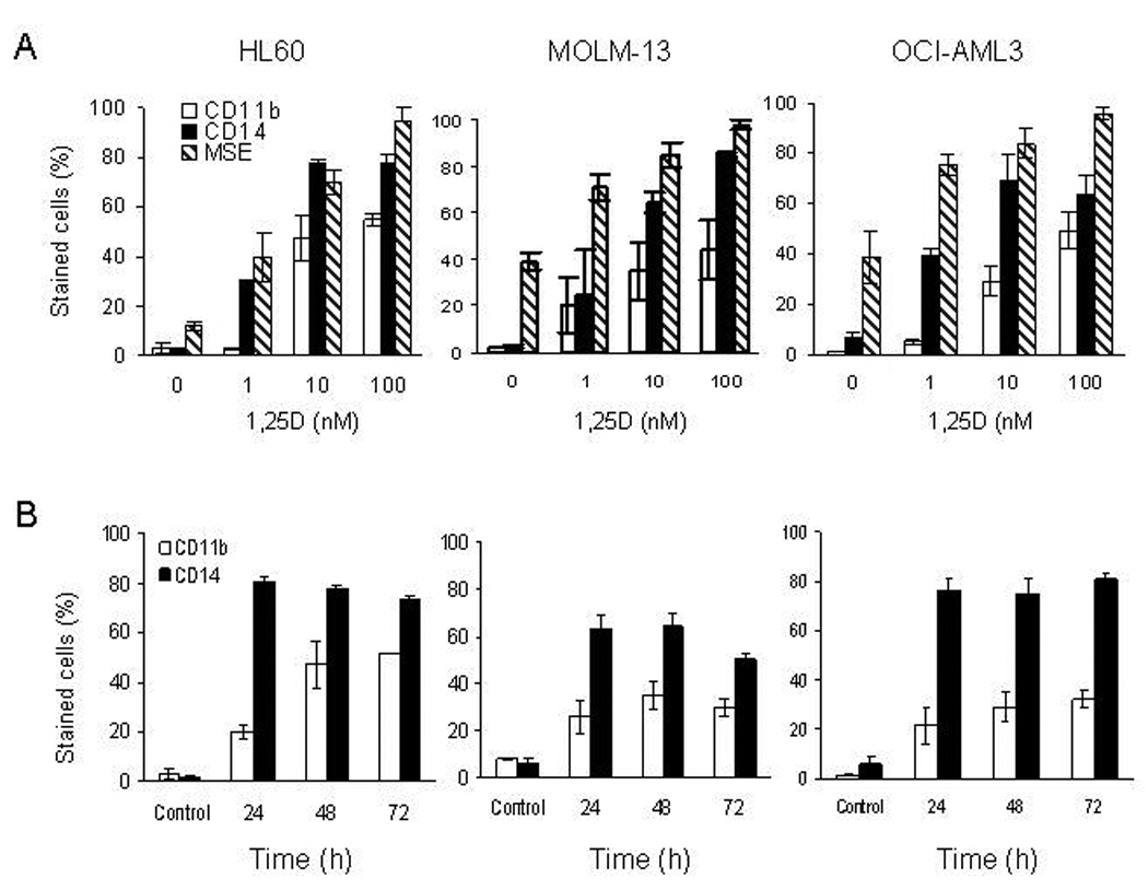
1,25D induces monocytic differentiation in p53-expressing and p53-null AML cells. A. Exponentially growing HL60, MOLM-13 and OCI-AML3 cells were exposed to 1, 10 and 100 nM 1,25D for 48 h. B. All three cell-lines were exposed to 10 nM 1,25D at the stated time. Treated cells were stained with anti-CD11b-FITC and anti-CD14-PE and assessed for surface expression by flow cytometry. Duplicate samples of treated cells were also stained for cytoplasmic monocyte specific esterase (MSE) and quantified by microscopy. The percentage of MSE-positive cells was determined by counting 100 cells in triplicate. The bars represent average values ± SD of three independent experiments, with duplicate data points.