
Figure 3.
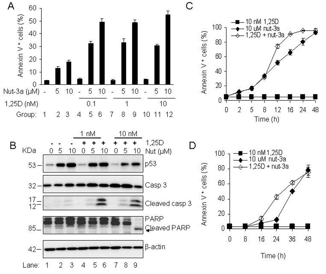
1,25D accelerates the onset of apoptosis by nutlin-3a in AML cells. A. OCI-AML3 cells were exposed to 0.1, 1 and 10 nM 1,25D for 48 h and nutlin-3a (5 and 10 µM) was added during the last 24 h. Cells were also exposed to single agents; 1,25D (0.1, 1, 10 nM) for 48 h and nutlin-3a (5, 10 µM) for 24 h. The percentage of Annexin V- positive cells was assessed with a Guava Nexin™ Kit. The bars represent average values ± SD of two independent experiments, with duplicate data points. Comparison of group 2 with groups 5, 8 and 11, and group 3 with groups 6, 9 and 12 all showed significant differences with p<0.05. P values were determined by Student t-test. B. OCI-AML3 cells were incubated with 1 and 10 nM 1,25D for 48 h and 5 µM or 10 µM nutlin-3a was added during the last 24 h. Cells were also incubated with single agents; 1,25D (1, 10 nM) for 48 h and nutlin-3a (5, 10 µM) for 24 h. The relative protein levels of p53, caspase-3 and PARP in whole cells lysates were analyzed by Western blotting, with β-actin as the loading protein. C and D. OCI-AML3 and MOLM-13 cells were incubated with 10 nM 1,25D for 48 h and 10 µM nutlin-3a was added during the last 2–48 h. Annexin V-positive cell fraction was determined with the Guava Nexin™ Kit. The bars represent average values ± SD of three independent experiments, with duplicate data points.