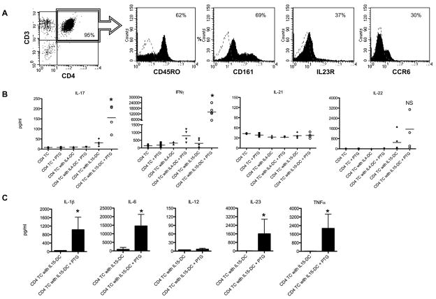
Figure 3. IL15-DC induce IL-17 and IFNγ secretion in cultures with autologous CD4+ T cells and PTG.
A, CD4+ T cells were purified by magnetic cell separation and analyzed by flow cytometry for the presence of Th17 surface markers. Dashed histograms are isotype controls and numbers are % positive cells. Histograms from 1 of 4 independent experiments are shown. B, CD4+ T cells were cultured alone or together with autologous IL15-DC or IL4-DC with and without 100μg/ml PTG for 72h. Cell-free supernatants were tested for secreted IL-17, IFNγ IL-21 and IL-22 by ELISA. Data are the means of 4–5 different donors tested in duplicate. C, The levels of IL-1β, IL-6, IL-12, IL-23 and TNFα were analyzed in the culture fluids from IL15-DC and CD4+ T cells incubated with and without 100μg/ml PTG for 72h by ELISA. Shown are the means of 4 independent experiments tested in duplicate. *, p<0.05 between T cell cultures with IL15-DC +/− PTG

