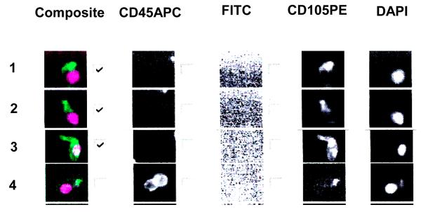
Figure 1.
This figure is representative of thumbnails of circulating endothelial cell candidates from a blood sample. From right to left the columns show the DAPI, CD105 PE, FITC, CD45 APC staining and a composite of DAPI (purple) and CD105 (green) staining. In order to be considered an endothelial cell the image should be CD105+, DAPI+, CD45−, FITC−. Rows 1, 2, 3, show endothelial cell staining with DAPI and CD105 but lacking CD45. Row number 4 shows leucocyte staining with DAPI, CD105 and CD45. The checks in the boxes indicate endothelial cell type and are tabulated by the software.

