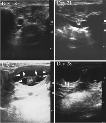
Fig. 1.
Ultrasonograms of the extra-fetal structures in pregnant Miniature Schnauzer bitches. Day 18: Transverse image of the first detection of an anechoic gestational sac. Day 21: Longitudinal image of the gestational sac. An echogenic inner placental layer was detected in the uterine wall. Day 27: Longitudinal image of the gestational sac contained an embryo and the tubular shape of the yolk sac membrane (white arrows). The zonary placenta (white arrowheads) was cylindrical in shape and appeared folded inward at the edges. Day 28: The amnionic membrane (white arrows) appeared faint.

