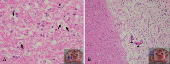
Fig. 4.
Microscopic features of the brain in an experimentally embolized dog (ID 2). (A) Thalamic lesion. Necrotic neurons (arrows), nuclear pyknosis, eosinophilia of the cytoplasm, and karyolysis were prominent. H&E stain, ×400. (B) Cortex lesion. Loss of tissue cohesion, infiltration by leukocytes (especially polymorphonuclear leukocytes), congestion of small parenchymal blood vessels (arrow heads), and angioblastic proliferation were observed. H&E stain, ×100.

