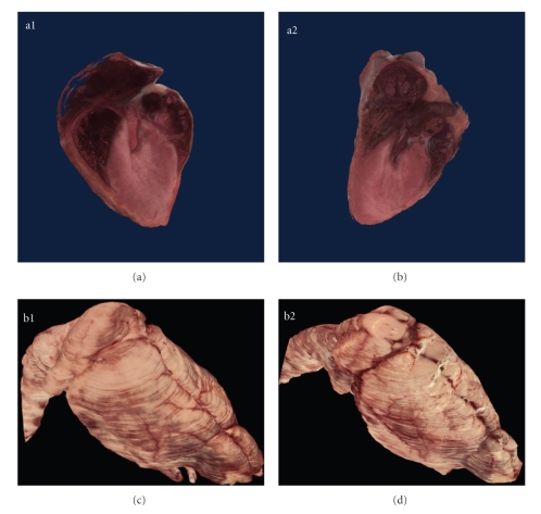
Figure 4.
Organ visualization. Cutaway through the 3D reconstructed hearts of the PEPCK-Cmus (a1) and control mouse (a2) reveals the chambers of the heart, pulmonary trunk and pulmonary veins. Manual segmentation of the heart from the 2D images also enabled computation of the heart volume. The heart of the PEPCK-Cmus mouse is 351.52 cubic mm in volume, while the heart from the control heart animal is 366.87 cubic mm. Other organs such as the brain of the PEPCK-Cmus (b1) and the control mouse (b2) were also semiautomatically segmented, visualized in 3D, and volumes computed. Table 1 lists the comparative volumes. The brain volume was also used as a normalizing factor for comparison of other organ volume estimates.

