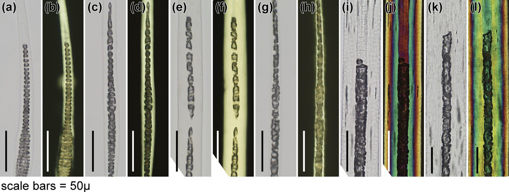
Figure 3.
Histology of specialized hairs. (a,c,e,g,i,k) White and (b,d,f,h,j,l) polarized microscopy revealed very subtle differences in septation patterns in the centre of(a–d) cilia (eyelashes), (e–h) tail hairs, and (i–l) vibrissae. Left pair, BALB/cByJ+/+; rght pair AKR/J-hid/hid mice (bar = 50 µm).

