
Figure 2.
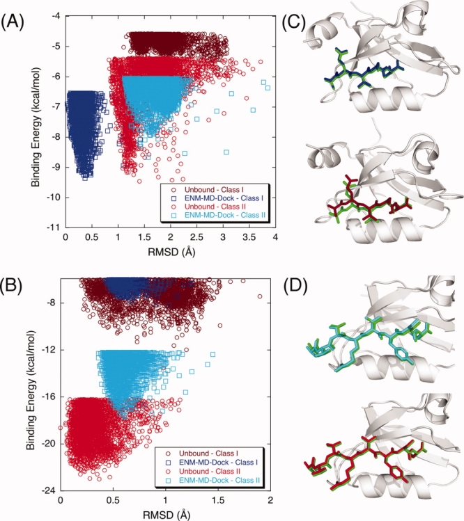
The binding energy score versus RMSD of the docked complex for (A) PSD-95 and (B) GRIP. Docking Class I and Class II peptides to the unbound conformation of PSD-95 does not discriminate the selectivity preference [brown (Class I) and red (Class II) dots in the plots]. However, when ENM-REMD snapshots are used (i.e., when the backbone flexibility is also considered), our flexible scheme does a better job than simply docking to the unbound structure of PSD-95 [blue (Class I) and cyan (Class II) dots in the plots]. (C) The comparison of docking Class I peptide to the unbound structure (upper figure) and the ENM-REMD snapshot (lower figure) is shown as ribbon diagrams. Green represents the actual binding mode in both docking, whereas blue and brown ones are for the docked peptide conformations corresponding to the lowest binding energies of ENM-REMD snapshot and the unbound structure, respectively. (D) Ribbon diagrams for docking Class II type of peptide to the unbound conformation and ENM-REMD conformation by RosettaLigand. [Color figure can be viewed in the online issue, which is available at http://www.interscience.wiley.com.]