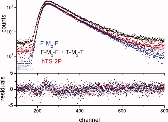
Figure 4.

Single-photon counting emission time profiles (top, λex = 480 nm, λem = 525 nm) and corresponding residues (bottom) of an equimolar mixture of F-M2-F and T-M2-T (black), of the dilabeled protein (hTS-2P, red) and of the F labeled TS (F-M2-F, blue). 1 channel corresponds to 51 ps. [Color figure can be viewed in the online issue, which is available at http://www.interscience.wiley.com.]
