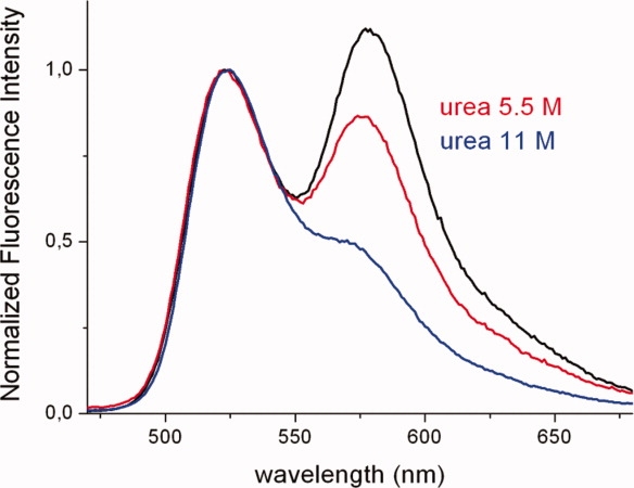
Figure 5.

Emission spectra of hTS-2P exposed for 3 h to urea at concentrations 0M (black), 5.5M (red), and 11M (blue). All spectra have been normalized at the F emission maximum (524 nm) for ease of comparison. [Color figure can be viewed in the online issue, which is available at http://www.interscience.wiley.com.]
