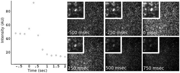
Fig. 5.
SynaptopHluorin vesicles (synaptic vesicles labeled with a pH sensitive green fluorescent protein) settled on the surface of a microchannel. At time 0 in the graph, we shifted a laminar boundary across the adsorbed vesicles. The vesicles were rapidly exchanged into a solution of SDS and NaOH. The intensity trace from the vesicles is shown on the left, and representative frames from the image sequence are shown on the right. As the pHluorin molecules come into contact with the alkaline solution they de-quench and brighten, followed shortly by the vesicle being disrupted and removed, resulting in a loss of fluorescence.

