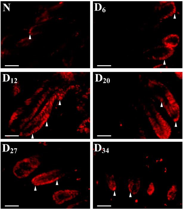
Figure 2. Crypt hyperplasia as measured by proliferating cell nuclear antigen (PCNA) staining.
Immunofluorescent labeling of PCNA as a marker of proliferation (arrow heads) in frozen sections prepared from non-infected (N) and infected (Days 6–34) mice. PCNA labeling correlated with changes in crypt hyperplasia during the time course of TMCH. Bar = 75 μm (n=5).

