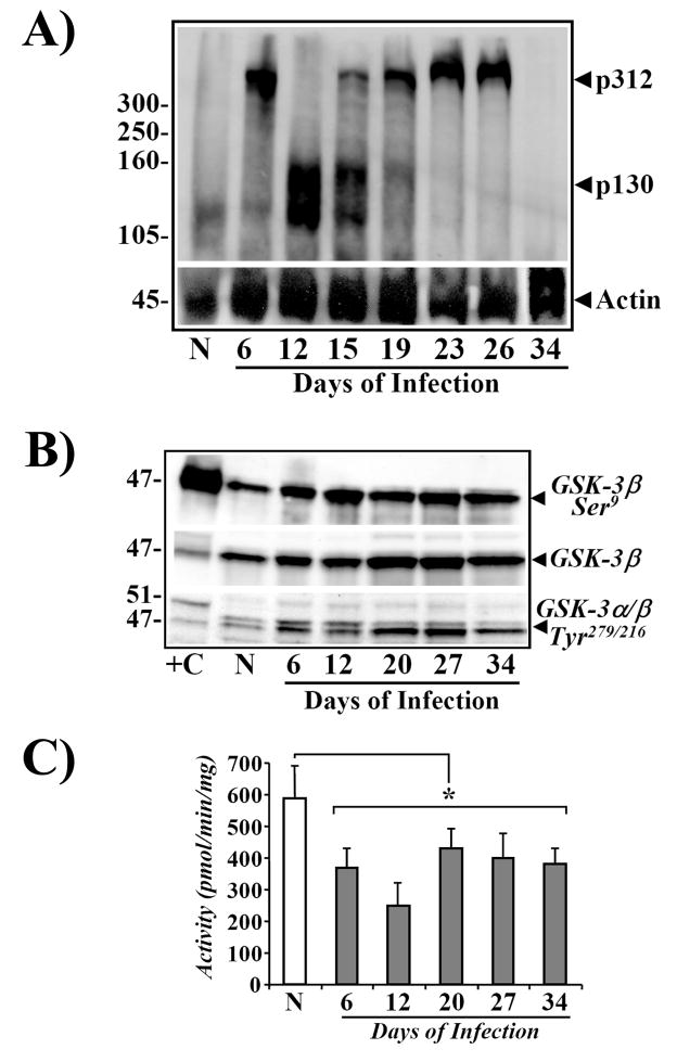
Figure 7.
A. Increases in cellular levels of APC during TMCH. Western blots showing relative levels of APC in non-infected (N) and Days 6–34 post-infected mouse distal colonic crypt samples. Triton X-100-solubilized total crypt extracts were analyzed by blotting with anti-APC N-terminal antibody.25 Protein loading was normalized to β-Actin (n=6). B. Phosphorylation status of GSK-3α/β during TMCH. Western blots showing relative levels of phosphorylated and total GSK-3α/β in non-infected (N) and Days 6–34 post-infected mouse distal colonic crypt samples. Triton X-100-solubilized total crypt extracts were analyzed by blotting with antibodies detecting either Ser-9-phosphorylated (GSK-3β-Ser9) or Tyr-279/216-phosphorylated GSK-3α/β and total GSK-3β. +C, mouse brain extract used as positive control. C. Measurement of GSK-3β activity during TMCH. Crypt cellular extracts from N and Days6–34 post-infected colons were immunoprecipitated with anti-GSK-3β and activity was measured in the immune-complexes with phospho-GSK-3β substrate. *P<0.05 over control; n = 2.

