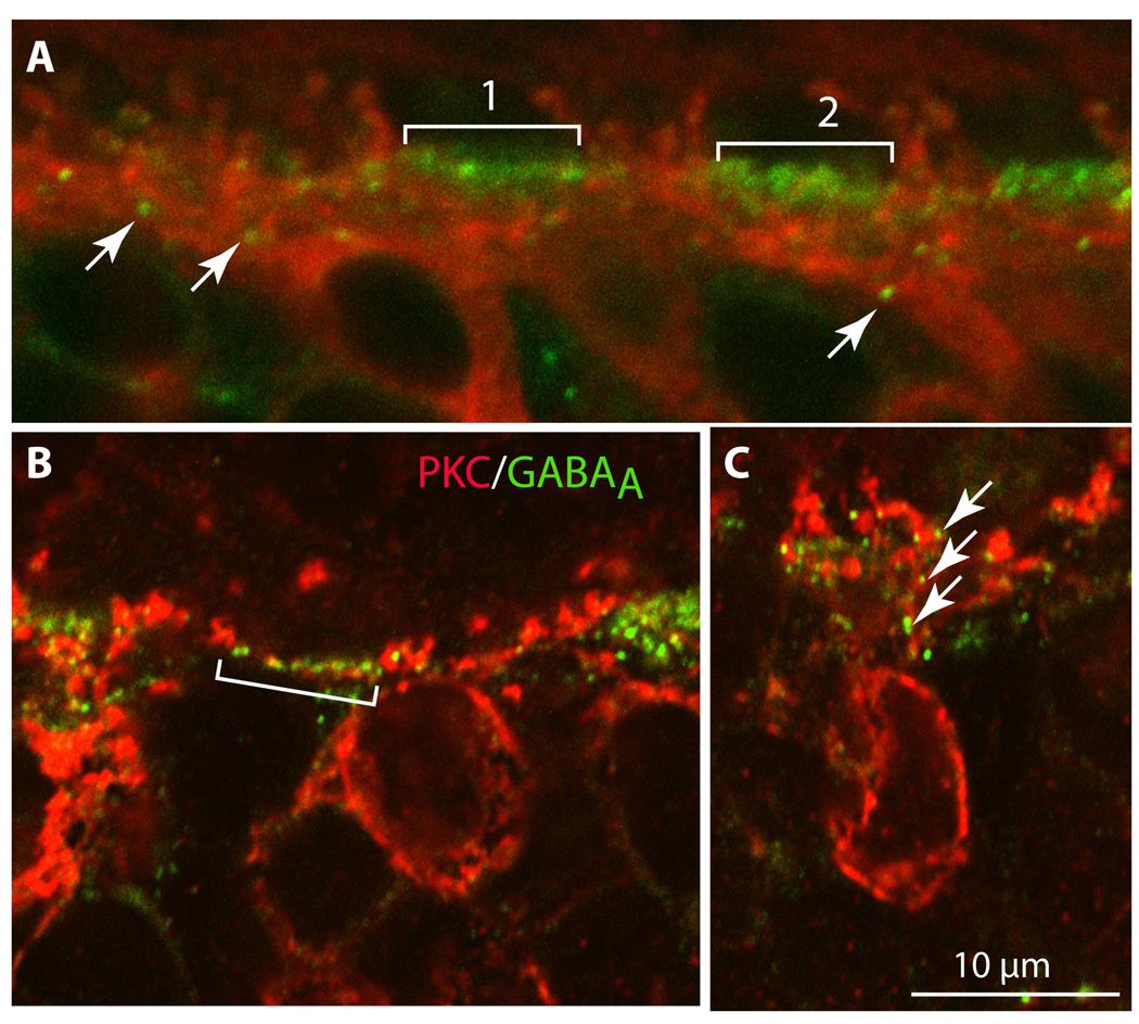
Figure 11. GABAA receptors are positioned in close proximity to PKC-stained dendrites in the OPL.
Double staining for PKC (red) and the α1 subunit of the GABAA receptor (green). In the first bracket in A, a string of GABAA receptor puncta (green) is flanked by red ascending rod bipolar dendrites that do not come in close proximity to them. In the second bracket, it is possible to see some red dendrites threading their way between the green puncta; these may be the dendrites of DB4 or other ON cone bipolar cells. In B, GABAA puncta (green in the bracket) are assembled in a row and are interspersed with red dots that appear to be a laterally ascending bipolar cell dendrite. In C, GABAA puncta (arrows) lie in close proximity to an ascending rod bipolar dendrite (imaged under 60x zoom 3).

