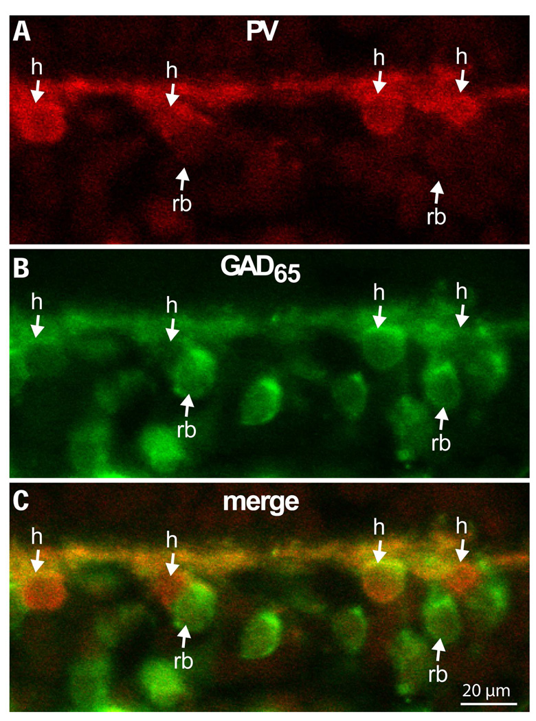
Figure 4. At mid retina, GAD65 staining in rod bipolar cells appears stronger than in horizontal cells.
Double labeling for parvalbumin (red, marks horizontal cells) and GAD65 (green) at 7 mm eccentricity. Down arrows point to horizontal cell somas (h) while up arrows point to rod bipolar somas (rb). Of the four horizontal cell somas shown in the figure, three stain more weakly for GAD65 than the adjacent rod bipolar cell somas (imaged under 40x).

