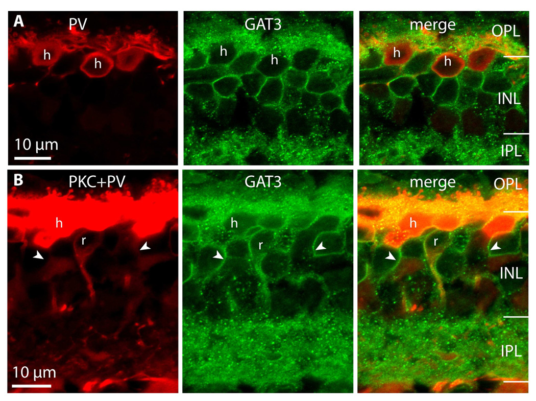
Figure 9. Horizontal cells express GAT3.
A. Double labeling for parvalbumin (PV, red) and GAT3 (green). Horizontal cell somas (h) stain for GAT3. (Imaged under 100x)
B. Triple staining for PKC+PV (red) and GAT3 (green). GAT3 labels both horizontal cells (h) and rod bipolar cells (r), but certain GAT3-positive cells belong to neither of these cell types (arrowhead lies inside the cell and points to the stained membrane) (imaged under 10x zoom 2).

