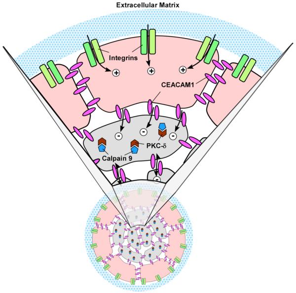
Figure 8. Model of CEACAM1 mediated lumen formation.
The peripheral acinar cells (beige) receive a life signal (plus sign) from ECM (blue mesh) via their interaction with cell surface integrins (green), while the central acinar cells (grey) do not receive this life signal. Instead the central acinar cells receive an apoptotic signal from CEACAM1 (red, minus sign). The apoptotic signal induces CAPN9 (brown) that cleaves PKC-δ (blue), that in turn, initiates apoptosis. Neither CEACAM1 nor CAPN9 are able to overcome the life signal produced by the ECM-integrin interaction in the peripheral acinar cells. Note: In the absence of CEACAM1, the central acinar cells do not die, CAPN9 is not induced, and PKC-δ is not cleaved.

