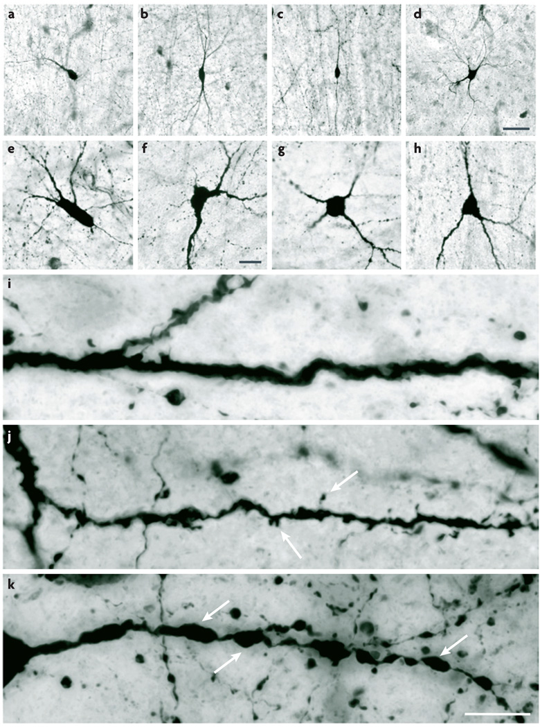
Figure 1. Somatic and dendritic morphology: shape and fine structure.
Human cerebral cortex sections stained by NADPH-diaphorase histochemistry reveal key features of somatic and dendritic morphology. a–h | Examples of soma shape and dendritic arborization polarity. Somata can be fusiform (a–c), polygonal (d), round (g), triangular (h) or shapes that are not described by any of these terms (e,f). Dendritic arborization can be termed monopolar (a), bitufted (b), bipolar (c) or multipolar (d). i–k | Higher-magnification images showing structural details of the dendrites. Dendrites can be fairly regular and aspiny (i), irregular and spiny (j; arrows indicate spines) or beaded and aspiny (k; arrows indicate beads). Scale bars: a–d (shown in d): 50 µm; e–h (shown in f): 20 µm; i–k (shown in k): 10 µm. Images supplied by Javier DeFelipe.

