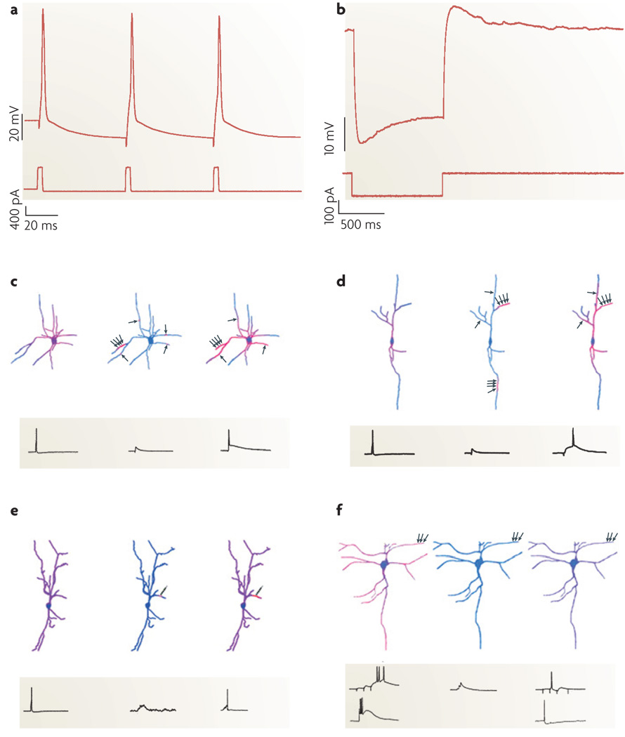
Figure 4. Functional characterization of interneurons.
a,b | Electrophysiological protocols used to measure neurons’ after hyperpolarization (AHP) and sag responses. Somatic current injection (bottom trace in both plots) and recorded membrane voltage (top traces) reveal an AHP (a) and a sag response (b)109. c–f | Diverse interneuron subclasses translate distinct patterns of activity into unique Ca2+ signals. The warmth of the colour reflects the magnitude of the Ca2+ accumulations. Axon terminals (indicated by the small black arrows) are positioned adjacent to dendrites to illustrate the spatial arrangement of synaptic activation, which can be in clusters or alone. c | A schematic of dendritic Ca2+ dynamics during a single action potential (left), a subthreshold excitatory postsynaptic potential (EPSP) (centre) and EPSP/action-potential coupling (right) in the dendrites of a multipolar perisoma-targeting fast-spiking cell from neocortical layer 2/3. Note that Ca2+ influx during single action potentials is proximally restricted. During subthreshold synaptic activation, convergent inputs activate dendritic subunits whereas individual inputs cause microdomains of Ca2+ accumulation. During EPSP/action-potential coupling, spikes propagate specifically to synaptically activated synaptic compartments where IA currents are inactivated. d | A schematic of a neocortical layer-2/3 calretinin-positive irregular-spiking interneuron stimulated as in c. The Ca2+ dynamics of bipolar irregular-spiking cells resemble fast-spiking cells, except that they lack Ca2+ microdomains. e | Dendritic Ca2+ signals in a layer-2/3 bitufted cell during action-potential backpropagation (left) and repetitive subthreshold (centre) and suprathreshold (right) activation of a single synapse. The multiple arrowheads on the individual schematic axonal terminals represent repetitive activation of a single synapse. f | Dendritic Ca2+ signals in a layer-5 rebound-spiking dendrite-targeting interneuron. In burst mode (left), synaptic activation (top trace) or somatic current injection (bottom trace) generates rebound spikes and highest Ca2+ signals at distal dendrites, where low-voltage-activated Ca2+ channels are clustered. In tonic mode (right), globally propagating Na+-based action potentials triggered either synaptically (top trace) or by somatic current injection (bottom trace) cause uniform Ca2+ accumulations. Excitation that is subthreshold for rebound-spike or action-potential generation does not cause Ca2+ influx (centre). Parts a and b reproduced from REF. 109. Parts c–f reproduced, with permission, from REF. 110 © (2005) Elsevier Science.

