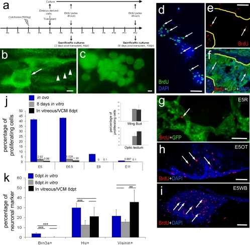
Figure 3.
Cells derived from embryonic retina, OT, and WB remain as masses of cells within the vitreous. (a) Injection and harvesting paradigm for experiments involving colchicine-induced retinal damage and transplantation of cells from GFP-transgenic embryos. (b) Representative micrograph of transplanted cells (arrow) adherent to the pecten (arrowheads). (c) Representative micrograph of aggregates of transplanted cells in the vitreous. Numbers of proliferating retinal cells significantly decreased after transplantation into the vitreous. (d–i) Representative micrographs of proliferating cells exposed to BrdU for 4 hours immediately before fixation. Cells were labeled with DAPI (blue) and with antibodies to GFP (green) and BrdU (red). (d) Cultured E7 retinal cells. (e, f) Sections through an aggregation of transplanted retinal cells adherent to the pecten. Arrows: BrdU+ cells. Yellow lines: border between the transplanted cells and the pecten. (g) Section through a VCM from transplanted E7 retinal cells. (h) Section through a VCM of transplanted E5 optic tectal cells. (i) Section through a VCM of transplanted E5 WB cells. (j) Histogram of the percentages of cells that incorporate BrdU in the vitreous after transplantation (black), in vitro (gray), and in ovo (blue). The percentage of proliferating cells is represented on the y-axis, and the developmental stage of the cells is represented on the x-axis. (j, insets) Percentage of proliferating cells from WB and OT. (k) Histogram of the percentages of neuronal markers in culture compared with transplanted cells within the VCM. Scale bars, 50 μm. **P ≤ 0.01; ***P ≤ 0.001.

