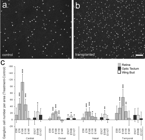
Figure 5.
Transplanted embryonic retinal cells promote the survival of ganglion cells in colchicine-treated eyes. (a, b) Representative confocal micrographs of Brn3a+ ganglion cells in central regions of colchicine-treated retinas from eyes that received vehicle (a) or transplants of E10 retinal cells (b). (c) Histogram illustrating the difference (mean ± SD for treated − control) in the numbers of ganglion cells per 0.45 mm2 of retinas from eyes that received control injections and those that received transplants. The source of the transplanted cells included the embryonic retina (gray bars), OT (black bars), and WB (white bars). The developmental stage at which the donor cells were harvested and the region of the retina in which cells were counted are indicated along the x-axis. *P ≤ 0.05; **P ≤ 0.01; ***P ≤ 0.001.

