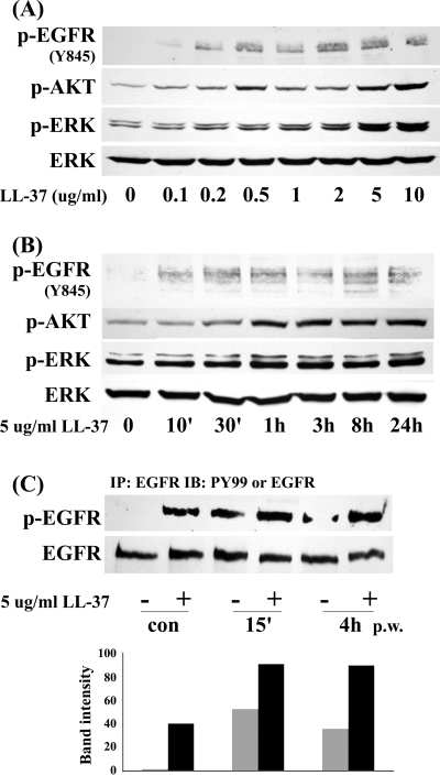
Figure 2.
LL-37 activates and prolongs EGFR signaling in HCECs. (A) Growth factor–starved THCE cells were stimulated with different concentrations of LL-37 (0.1 μg/mL to 10 μg/mL) for 1 hour. (B) Growth factor–starved primary HCECs were stimulated with 5 μg/mL LL-37 for various time points (10 minutes to 24 hours). Cells were then lysed and subjected to Western blot analysis with anti–phospho-EGFR (Y845), phospho-AKT1/2 (p-AKT), phospho-ERK1/2 (p-ERK), and ERK2 (as a loading control) antibodies. (C) THCE cells were stimulated with extensive wounding, 5 μg/mL LL-37, or both for 15 minutes or 4 hours before they were lysed. THCE lysates were immunoprecipitated with EGFR antibody, immunoblotted with anti–PY99 antibody (p-EGFR), and reprobed with EGFR antibody (EGFR) to access the amount of EGFR precipitated. Bar graph represents the average band densities of p-EGFR normalized against total EGFR from two independent experiments.

